The appendix is a small, tube-like organ attached to the large intestine. Inside the appendix, there is special tissue called lymphoid tissue, which is part of the immune system. This tissue helps fight off infections.
The vermiform appendix is a small, worm-shaped organ attached to the large intestine on the right side of the lower abdomen. Despite its small size, it plays a significant role in the immune system due to its rich content of lymphoid tissue, which helps protect against infections in the intestines.

When the body detects harmful substances like viruses and bacteria in the peritoneal cavity (the space within the abdomen that houses the intestines, liver, and other organs), the lymphoid tissue in the appendix responds by swelling and expanding. This reaction is part of the body’s immune response to fight off these invaders.
As the lymphoid tissue swells, the appendix can extend its length and change its position slightly to better reach and absorb harmful materials present in the peritoneal cavity. This includes free peritoneal fluid, deteriorated tissue, and retrograde menstrual blood (menstrual blood that flows backward into the peritoneal cavity instead of exiting the body).
By absorbing these substances, the appendix helps to clear the peritoneal cavity of potentially harmful materials, aiding in the body’s overall defense mechanisms. The lymphoid cells within the appendix work to neutralize and eliminate pathogens, thereby contributing to the immune protection of the abdominal region. This function has led to the appendix being referred to as the “abdominal tonsil,” similar to the role of tonsils in protecting the throat from infection.
Position of the Appendix
Variations in the position include retrocecal(but intraperitoneal), subcecal, pre-ileal and post-ileal, pelvic and as far as into the hepatorenal recess. In addition, factors such as posture, respiration, and distention of adjacent bowels can influence the position of the appendix. In addition, factors such as posture, respiration, and distention of adjacent bowels can influence the position of the appendix. 1Hodge BD, Kashyap S, Khorasani-Zadeh A. Anatomy, Abdomen and Pelvis: Appendix. [Updated 2023 Aug 8]. In: StatPearls [Internet]. Treasure Island (FL): StatPearls Publishing; 2024 Jan-.

Length and Diameter of Appendix
The appendix is subject to the extremes of variation. Its length varies from 2 to 20 cm, in average 9 cm.2Singh IB. Human Anatomy, Regional and Applied. 3rd edition. Vol. 2. New Delhi, India: CBC Publishers and Distributors; 1999. The base of appendix is connected to the cecum, but its head can be placed in different situations. The diversity of situations is categorized into six locations: retrocecal, pelvic, subcecal, preileal, retroileal, and ectopic.3Schwartz SJ, Shires GT, Spencer FC, Daly JM, Fischer JE, Galloway AC. The Appendix. 7th edition. Philadelphia, Pa, USA: McGraw-Hill; 1999. Principles of surgery Schwartz; pp. 1383–1385., 4Sabiston DC, Courtney M. Sabiston’s Textbook of Surgery: The Biological Basis of Modern Surgical Practice in Appendix. 16th edition. Vol. 2. Philadelphia, Pa, USA: WB Saunders; 2001., 5Williams PL, Bannister LH, Berry MM, Collins P, Dyson M, Dussek JE. Alimentary System. 39th edition. New York, NY, USA: Churchill Livingstone; 2005. Gray’s anatomy; pp. 1775–1776., 6Nayak BS. Why the tip of vermiform appendix has variable position? Medical Hypotheses. 2010;75(6):682–683. Ghorbani A, Forouzesh M, Kazemifar AM. Variation in Anatomical Position of Vermiform Appendix among Iranian Population: An Old Issue Which Has Not Lost Its Importance. Anat Res Int. 2014;2014:313575.
The diameter of the normal appendix is 6 mm, with a normal range encompassing 3–9 mm.7Trout AT, Towbin AJ, Zhang B. Journal club: The pediatric appendix: defining normal. AJR Am J Roentgenol. 2014 May;202(5):936-45. doi: 10.2214/AJR.13.11030. PMID: 24758645.
Appendicitis should not be diagnosed by size alone. Normal appendices can measure 13 mm in width and 35 cm in length, so it is important to consider the additional findings of obstruction, ischemia, inflammation and perforation. Obstruction is highly associated with perforation and complications whereas cases of ‘simple’ non-obstructive appendicitis may have a benign course, resolving spontaneously. These cases may be caused by retrograde menstrual blood and endometrial tissue in women.8Orscheln E & Trout A. Appendiceal Diameter: CT Versus Sonographic Measurements. Pediatr Radiol. 2016;46(3):316-21., 9Noguchi T, Yoshimitsu K, Yoshida M. Periappendiceal Hyperechoic Structure on Sonography: A Sign of Severe Appendicitis. J Ultrasound Med. 2005;24(3):323-7, 10Rettenbacher T, Hollerweger A, Macheiner P et al. Outer Diameter of the Vermiform Appendix as a Sign of Acute Appendicitis: Evaluation at US. Radiology. 2001;218(3):757-62., 11Ives E, Sung S, McCue P, Durrani H, Halpern E. Independent Predictors of Acute Appendicitis on CT with Pathologic Correlation. Acad Radiol. 2008;15(8):996-1003., 12Willekens I, Peeters E, De Maeseneer M, de Mey J. The Normal Appendix on CT: Does Size Matter? PLoS One. 2014;9(5):e96476., 13Orscheln E & Trout A. Appendiceal Diameter: CT Versus Sonographic Measurements. Pediatr Radiol. 2016;46(3):316-21., 14Cho J, Akers M, Siavoshi M, Gress T, Thompson E. Features on Computed Tomography That Correlate With Acute Appendicitis. Am Surg. 2023;89(6):2876-9.,15Park NH, Oh HE, Park HJ, Park JY. Ultrasonography of normal and abnormal appendix in children. World J Radiol. 2011 Apr 28;3(4):85-91.


How Does the Lymphoid Tissue Work?
- Swelling and Expansion: When the body detects something harmful or unusual, like deteriorated tissue or fluids that shouldn’t be there, the lymphoid tissue in the appendix can swell and expand. This is similar to how your lymph nodes swell when you have a cold.
- Extending its Length and Changing Position: As the lymphoid tissue swells, the appendix itself can stretch and change its position slightly. This movement helps it to reach and absorb different substances in the abdominal cavity.

Absorption Process:
- Free Peritoneal Fluid: The abdomen (belly area) has a thin layer of fluid called peritoneal fluid that helps lubricate the organs. If there’s excess fluid, the appendix can absorb some of it.
- Pancreatic Juices and Enzymes: Digestive enzymes, and pancreatic juices that leak from the pancreas pseudocysts during conditions like pancreatitis. The appendix can help absorb these enzymes and pancreatic juices that mix with the Free Peritoneal Fluid to prevent them from causing damage elsewhere.
- Deteriorated Tissue: Sometimes, old or damaged tissues are present in the abdomen. The appendix can help in absorbing and removing these unwanted tissues.
- Retrograde Menstrual Blood and Endometrial Tissue: Occasionally, menstrual blood and tissue can move backward into the abdomen instead of leaving the body. The appendix helps to absorb and remove this tissue, reducing irritation and potential infections.

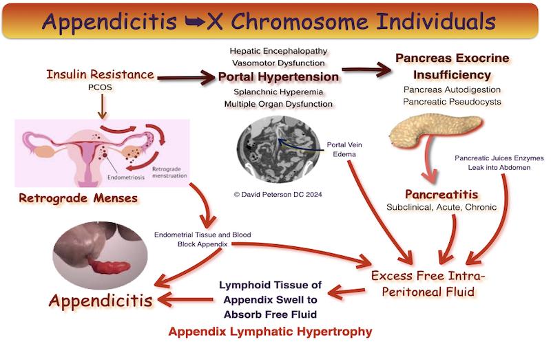

The appendix can become overwhelmed when absorbing excess free peritoneal fluid (fluid in the abdominal cavity), digestive enzymes, pancreatic juices that leak from the pancreas during conditions like pancreatitis in both men and women as well as retrograde menstrual blood and endometrial tissue that leak from the fallopian tube during menstruation.
Why is This Important?
The ability of the appendix to absorb these substances helps keep the abdomen clean and prevents infections or inflammation. It’s a part of the body’s way to manage waste and maintain a healthy environment inside the abdomen.
So, in simple terms, the appendix and its lymphoid tissue swell and stretch to reach and absorb harmful fluids and tissues in the abdomen, helping to keep the belly area clean and free of infection.
References
- 1Hodge BD, Kashyap S, Khorasani-Zadeh A. Anatomy, Abdomen and Pelvis: Appendix. [Updated 2023 Aug 8]. In: StatPearls [Internet]. Treasure Island (FL): StatPearls Publishing; 2024 Jan-.
- 2Singh IB. Human Anatomy, Regional and Applied. 3rd edition. Vol. 2. New Delhi, India: CBC Publishers and Distributors; 1999.
- 3Schwartz SJ, Shires GT, Spencer FC, Daly JM, Fischer JE, Galloway AC. The Appendix. 7th edition. Philadelphia, Pa, USA: McGraw-Hill; 1999. Principles of surgery Schwartz; pp. 1383–1385.
- 4Sabiston DC, Courtney M. Sabiston’s Textbook of Surgery: The Biological Basis of Modern Surgical Practice in Appendix. 16th edition. Vol. 2. Philadelphia, Pa, USA: WB Saunders; 2001.
- 5Williams PL, Bannister LH, Berry MM, Collins P, Dyson M, Dussek JE. Alimentary System. 39th edition. New York, NY, USA: Churchill Livingstone; 2005. Gray’s anatomy; pp. 1775–1776.
- 6Nayak BS. Why the tip of vermiform appendix has variable position? Medical Hypotheses. 2010;75(6):682–683. Ghorbani A, Forouzesh M, Kazemifar AM. Variation in Anatomical Position of Vermiform Appendix among Iranian Population: An Old Issue Which Has Not Lost Its Importance. Anat Res Int. 2014;2014:313575.
- 7Trout AT, Towbin AJ, Zhang B. Journal club: The pediatric appendix: defining normal. AJR Am J Roentgenol. 2014 May;202(5):936-45. doi: 10.2214/AJR.13.11030. PMID: 24758645.
- 8Orscheln E & Trout A. Appendiceal Diameter: CT Versus Sonographic Measurements. Pediatr Radiol. 2016;46(3):316-21.
- 9Noguchi T, Yoshimitsu K, Yoshida M. Periappendiceal Hyperechoic Structure on Sonography: A Sign of Severe Appendicitis. J Ultrasound Med. 2005;24(3):323-7
- 10Rettenbacher T, Hollerweger A, Macheiner P et al. Outer Diameter of the Vermiform Appendix as a Sign of Acute Appendicitis: Evaluation at US. Radiology. 2001;218(3):757-62.
- 11Ives E, Sung S, McCue P, Durrani H, Halpern E. Independent Predictors of Acute Appendicitis on CT with Pathologic Correlation. Acad Radiol. 2008;15(8):996-1003.
- 12Willekens I, Peeters E, De Maeseneer M, de Mey J. The Normal Appendix on CT: Does Size Matter? PLoS One. 2014;9(5):e96476.
- 13Orscheln E & Trout A. Appendiceal Diameter: CT Versus Sonographic Measurements. Pediatr Radiol. 2016;46(3):316-21.
- 14Cho J, Akers M, Siavoshi M, Gress T, Thompson E. Features on Computed Tomography That Correlate With Acute Appendicitis. Am Surg. 2023;89(6):2876-9.
- 15Park NH, Oh HE, Park HJ, Park JY. Ultrasonography of normal and abnormal appendix in children. World J Radiol. 2011 Apr 28;3(4):85-91.

