Length and Diameter of Appendix
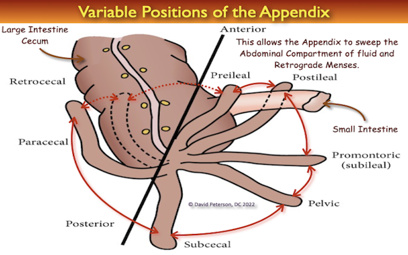
The appendix is subject to the extremes of variation. Its length varies from 2 to 20 cm, in average 9 cm.1Singh IB. Human Anatomy, Regional and Applied. 3rd edition. Vol. 2. New Delhi, India: CBC Publishers and Distributors; 1999. The base of appendix is connected to the cecum, but its head can be placed in different situations. The diversity of situations is categorized into six locations: retrocecal, pelvic, subcecal, preileal, retroileal, and ectopic.2Schwartz SJ, Shires GT, Spencer FC, Daly JM, Fischer JE, Galloway AC. The Appendix. 7th edition. Philadelphia, Pa, USA: McGraw-Hill; 1999. Principles of surgery Schwartz; pp. 1383–1385. 3Sabiston DC, Courtney M. Sabiston’s Textbook of Surgery: The Biological Basis of Modern Surgical Practice in Appendix. 16th edition. Vol. 2. Philadelphia, Pa, USA: WB Saunders; 2001. 4Williams PL, Bannister LH, Berry MM, Collins P, Dyson M, Dussek JE. Alimentary System. 39th edition. New York, NY, USA: Churchill Livingstone; 2005. Gray’s anatomy; pp. 1775–1776. 5Nayak BS. Why the tip of vermiform appendix has variable position? Medical Hypotheses. 2010;75(6):682–683. 6Ghorbani A, Forouzesh M, Kazemifar AM. Variation in Anatomical Position of Vermiform Appendix among Iranian Population: An Old Issue Which Has Not Lost Its Importance. Anat Res Int. 2014;2014:313575.
The diameter of the normal appendix was 6 mm, with a normal range encompassing 3–9 mm. 7Trout AT, Towbin AJ, Zhang B. Journal club: The pediatric appendix: defining normal. AJR Am J Roentgenol. 2014 May;202(5):936-45. doi: 10.2214/AJR.13.11030. PMID: 24758645.
Appendicitis should not be diagnosed by size alone. Normal appendices can measure 13 mm in width and 35 cm in length, so it is important to consider the additional findings of obstruction, ischemia, inflammation and perforation. Obstruction is highly associated with perforation and complications whereas cases of ‘simple’ non-obstructive appendicitis may have a benign course, resolving spontaneously. These cases may be caused by retrograde menstrual blood and endometrial tissue in women. 8Orscheln E & Trout A. Appendiceal Diameter: CT Versus Sonographic Measurements. Pediatr Radiol. 2016;46(3):316-21. 9Noguchi T, Yoshimitsu K, Yoshida M. Periappendiceal Hyperechoic Structure on Sonography: A Sign of Severe Appendicitis. J Ultrasound Med. 2005;24(3):323-7; 10Rettenbacher T, Hollerweger A, Macheiner P et al. Outer Diameter of the Vermiform Appendix as a Sign of Acute Appendicitis: Evaluation at US. Radiology. 2001;218(3):757-62. 11Ives E, Sung S, McCue P, Durrani H, Halpern E. Independent Predictors of Acute Appendicitis on CT with Pathologic Correlation. Acad Radiol. 2008;15(8):996-1003. 12Willekens I, Peeters E, De Maeseneer M, de Mey J. The Normal Appendix on CT: Does Size Matter? PLoS One. 2014;9(5):e96476. 13Orscheln E & Trout A. Appendiceal Diameter: CT Versus Sonographic Measurements. Pediatr Radiol. 2016;46(3):316-21. 14Cho J, Akers M, Siavoshi M, Gress T, Thompson E. Features on Computed Tomography That Correlate With Acute Appendicitis. Am Surg. 2023;89(6):2876-9. 15Park NH, Oh HE, Park HJ, Park JY. Ultrasonography of normal and abnormal appendix in children. World J Radiol. 2011 Apr 28;3(4):85-91.
Dive into the fascinating world of human anatomy with a focus on the appendix! This video explores the variability in the size of the appendix, from its typical dimensions to how it can change under different physiological conditions. We’ll discuss how conditions like lymphatic hypertrophy can affect its size, and explore the complexities of diagnosing appendix-related issues due to misleading symptoms caused by various abdominal and pelvic substances. Whether you’re a medical student, a healthcare professional, or just curious about human biology, this video will shed light on why diagnosing appendix problems can be more challenging than you think.
If you found this video informative, don’t forget to like, subscribe, and hit the bell icon for more deep dives into medical mysteries and health education. Share your thoughts or experiences in the comments below!
References
- 1Singh IB. Human Anatomy, Regional and Applied. 3rd edition. Vol. 2. New Delhi, India: CBC Publishers and Distributors; 1999.
- 2Schwartz SJ, Shires GT, Spencer FC, Daly JM, Fischer JE, Galloway AC. The Appendix. 7th edition. Philadelphia, Pa, USA: McGraw-Hill; 1999. Principles of surgery Schwartz; pp. 1383–1385.
- 3Sabiston DC, Courtney M. Sabiston’s Textbook of Surgery: The Biological Basis of Modern Surgical Practice in Appendix. 16th edition. Vol. 2. Philadelphia, Pa, USA: WB Saunders; 2001.
- 4Williams PL, Bannister LH, Berry MM, Collins P, Dyson M, Dussek JE. Alimentary System. 39th edition. New York, NY, USA: Churchill Livingstone; 2005. Gray’s anatomy; pp. 1775–1776.
- 5Nayak BS. Why the tip of vermiform appendix has variable position? Medical Hypotheses. 2010;75(6):682–683.
- 6Ghorbani A, Forouzesh M, Kazemifar AM. Variation in Anatomical Position of Vermiform Appendix among Iranian Population: An Old Issue Which Has Not Lost Its Importance. Anat Res Int. 2014;2014:313575.
- 7Trout AT, Towbin AJ, Zhang B. Journal club: The pediatric appendix: defining normal. AJR Am J Roentgenol. 2014 May;202(5):936-45. doi: 10.2214/AJR.13.11030. PMID: 24758645.
- 8Orscheln E & Trout A. Appendiceal Diameter: CT Versus Sonographic Measurements. Pediatr Radiol. 2016;46(3):316-21.
- 9Noguchi T, Yoshimitsu K, Yoshida M. Periappendiceal Hyperechoic Structure on Sonography: A Sign of Severe Appendicitis. J Ultrasound Med. 2005;24(3):323-7;
- 10Rettenbacher T, Hollerweger A, Macheiner P et al. Outer Diameter of the Vermiform Appendix as a Sign of Acute Appendicitis: Evaluation at US. Radiology. 2001;218(3):757-62.
- 11Ives E, Sung S, McCue P, Durrani H, Halpern E. Independent Predictors of Acute Appendicitis on CT with Pathologic Correlation. Acad Radiol. 2008;15(8):996-1003.
- 12Willekens I, Peeters E, De Maeseneer M, de Mey J. The Normal Appendix on CT: Does Size Matter? PLoS One. 2014;9(5):e96476.
- 13Orscheln E & Trout A. Appendiceal Diameter: CT Versus Sonographic Measurements. Pediatr Radiol. 2016;46(3):316-21.
- 14Cho J, Akers M, Siavoshi M, Gress T, Thompson E. Features on Computed Tomography That Correlate With Acute Appendicitis. Am Surg. 2023;89(6):2876-9.
- 15Park NH, Oh HE, Park HJ, Park JY. Ultrasonography of normal and abnormal appendix in children. World J Radiol. 2011 Apr 28;3(4):85-91.